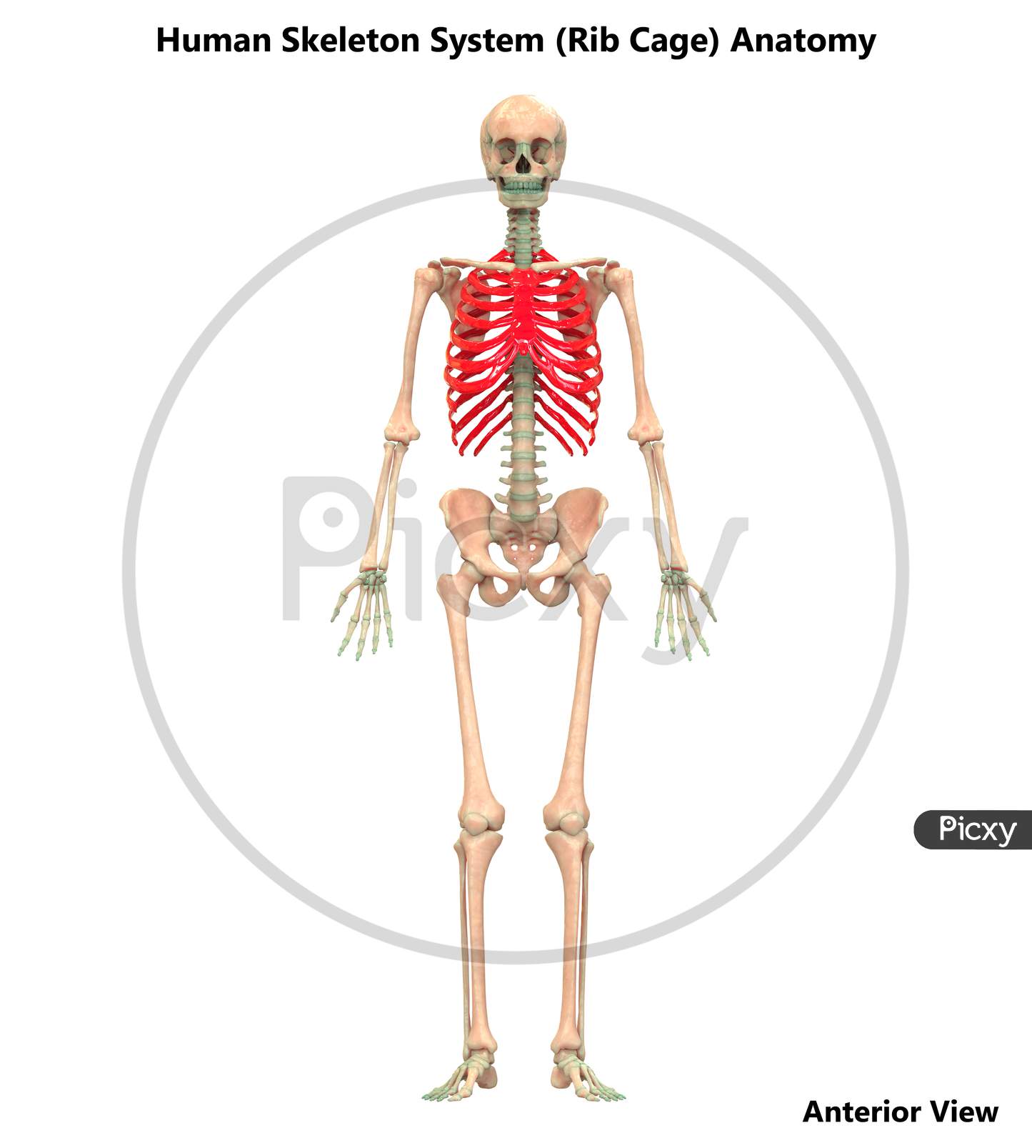
Anatomy Of Rib Cage : Image Of Human Skeleton System Anatomy Rib Cage Ci993068 Picxy / The thoracic cage takes the form of a domed bird cage with the horizontal bars formed by ribs and costal cartilages.
Anatomy Of Rib Cage : Image Of Human Skeleton System Anatomy Rib Cage Ci993068 Picxy / The thoracic cage takes the form of a domed bird cage with the horizontal bars formed by ribs and costal cartilages.. The ribs form the main structure of the thoracic cage that protects the thoracic organs. See facet joint anatomy animation. It is supported by the vertical sternum or. Feb 10, 2020 · anatomy. These joints are where a vertebra connects, or articulates, with a rib.
A cervical rib forms from the overdevelopment of the transverse process of a cervical vertebra, typically from the seventh cervical vertebra in the neck known as c7. It is made up of 12 pairs of ribs. The first rib is the most superior of the twelve ribs. An inhalation is accomplished when the muscular diaphragm, at the floor of the thoracic cavity, contracts and flattens, while the contraction of intercostal muscles lift the rib cage up and out. It has a roughened area on its upper surface, from which the serratus anterior muscle originates.
Image Of Human Skeleton System Anatomy Rib Cage Ci993068 Picxy from images.picxy.com The flank or latus is the side of the body between the rib cage and the iliac bone of the hip (below the rib cage and above the ilium). The human rib cage is a component of the human respiratory system. It is supported by the vertical sternum or. A cervical rib forms from the overdevelopment of the transverse process of a cervical vertebra, typically from the seventh cervical vertebra in the neck known as c7. See facet joint anatomy animation. Feb 10, 2020 · anatomy. Jul 29, 2021 · the thoracic cage (rib cage) is the skeleton of the thoracic wall. It encloses the thoracic cavity, which contains the lungs.
The rib cage is a bony structure found in the chest (thoracic cavity).
1 it is sometimes called the lumbar region. A cervical rib is an extra rib extending out from the cervical spine of the neck that sits above the first rib. Vertebrae surround and protect the spinal cord and bones of the rib cage help protect the heart and lungs of the thorax. Dec 21, 2020 · anatomy the rib cage has 12 sets of ribs. It is formed by the 12 thoracic vertebrae, 12 pairs of ribs and associated costal cartilages and the sternum. The human rib cage is a component of the human respiratory system. It presents as an additional rib coming off t13 or l1 (depending on numbering classification) and m. It is one of the borders of the superior thoracic aperture. There are 12 pairs of ribs which are separated by intercostal spaces. The thoracic cage takes the form of a domed bird cage with the horizontal bars formed by ribs and costal cartilages. These joints are where a vertebra connects, or articulates, with a rib. An inhalation is accomplished when the muscular diaphragm, at the floor of the thoracic cavity, contracts and flattens, while the contraction of intercostal muscles lift the rib cage up and out. It is supported by the vertical sternum or.
There are 12 pairs of ribs which are separated by intercostal spaces. The first rib is the most superior of the twelve ribs. The ribs form the main structure of the thoracic cage that protects the thoracic organs. See facet joint anatomy animation. The costocorporeal joint is where the rib head connects with two adjacent vertebral bodies and the disc between them.
Rib Cage Anatomy Function Britannica from cdn.britannica.com Bones work together with muscles as simple mechanical lever systems to produce body movement. The costocorporeal joint is where the rib head connects with two adjacent vertebral bodies and the disc between them. It is formed by the 12 thoracic vertebrae, 12 pairs of ribs and associated costal cartilages and the sternum. Feb 10, 2020 · anatomy. It is one of the borders of the superior thoracic aperture. Lumbar (or 13th) ribs are a rare anatomical variant and represent transitional vertebrae at the thoracolumbar junction with a prevalence of ~1% 1. The flank or latus is the side of the body between the rib cage and the iliac bone of the hip (below the rib cage and above the ilium). There are 12 pairs of ribs which are separated by intercostal spaces.
The human rib cage is a component of the human respiratory system.
Jul 29, 2021 · the thoracic cage (rib cage) is the skeleton of the thoracic wall. It presents as an additional rib coming off t13 or l1 (depending on numbering classification) and m. Lumbar (or 13th) ribs are a rare anatomical variant and represent transitional vertebrae at the thoracolumbar junction with a prevalence of ~1% 1. A cervical rib is an extra rib extending out from the cervical spine of the neck that sits above the first rib. An inhalation is accomplished when the muscular diaphragm, at the floor of the thoracic cavity, contracts and flattens, while the contraction of intercostal muscles lift the rib cage up and out. These joints are where a vertebra connects, or articulates, with a rib. Bones work together with muscles as simple mechanical lever systems to produce body movement. It encloses the thoracic cavity, which contains the lungs. A cervical rib forms from the overdevelopment of the transverse process of a cervical vertebra, typically from the seventh cervical vertebra in the neck known as c7. The first rib is the most superior of the twelve ribs. Rib 2 is thinner and longer than rib 1, and has two articular facets on the head as normal. 1 it is sometimes called the lumbar region. It has a roughened area on its upper surface, from which the serratus anterior muscle originates.
Dec 21, 2020 · anatomy the rib cage has 12 sets of ribs. The costocorporeal joint is where the rib head connects with two adjacent vertebral bodies and the disc between them. 1 it is sometimes called the lumbar region. Jul 29, 2021 · the thoracic cage (rib cage) is the skeleton of the thoracic wall. It presents as an additional rib coming off t13 or l1 (depending on numbering classification) and m.
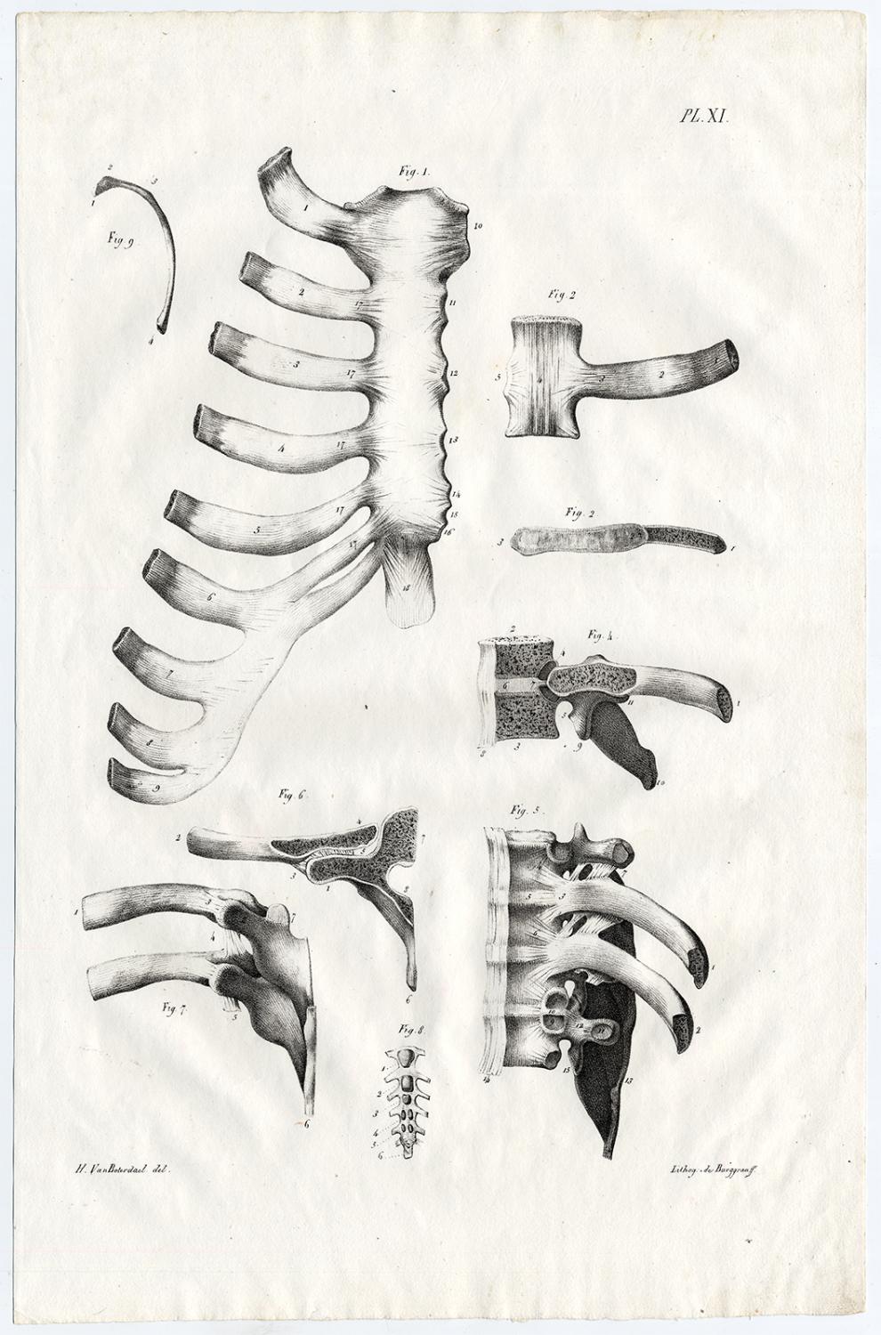
Antique Print Human Anatomy Rib Cage Bones Cloquet 1821 Kunst Nbsp Nbsp Grafik Nbsp Nbsp Poster Theprintscollector from pictures.abebooks.com It encloses the thoracic cavity, which contains the lungs. It is one of the borders of the superior thoracic aperture. The flank or latus is the side of the body between the rib cage and the iliac bone of the hip (below the rib cage and above the ilium). The thoracic cage takes the form of a domed bird cage with the horizontal bars formed by ribs and costal cartilages. The ribs form the main structure of the thoracic cage that protects the thoracic organs. Lumbar (or 13th) ribs are a rare anatomical variant and represent transitional vertebrae at the thoracolumbar junction with a prevalence of ~1% 1. 1 it is sometimes called the lumbar region. It is supported by the vertical sternum or.
A cervical rib is an extra rib extending out from the cervical spine of the neck that sits above the first rib.
It presents as an additional rib coming off t13 or l1 (depending on numbering classification) and m. The rib cage is a bony structure found in the chest (thoracic cavity). Dec 21, 2020 · anatomy the rib cage has 12 sets of ribs. An inhalation is accomplished when the muscular diaphragm, at the floor of the thoracic cavity, contracts and flattens, while the contraction of intercostal muscles lift the rib cage up and out. The ribs form the main structure of the thoracic cage that protects the thoracic organs. It has a roughened area on its upper surface, from which the serratus anterior muscle originates. 1 it is sometimes called the lumbar region. Feb 10, 2020 · anatomy. The costocorporeal joint is where the rib head connects with two adjacent vertebral bodies and the disc between them. It is formed by the 12 thoracic vertebrae, 12 pairs of ribs and associated costal cartilages and the sternum. The first rib is the most superior of the twelve ribs. It encloses the thoracic cavity, which contains the lungs. The thoracic cage takes the form of a domed bird cage with the horizontal bars formed by ribs and costal cartilages.


0 Komentar